Author: Jonathan Kobles, MD
Warning, this article contains graphic images.
Introduction:
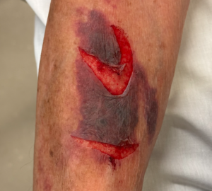
An 85-year-old male with a history of chronic lymphocytic leukemia (CLL) and atrial fibrillation on Eliquis presents to the emergency department (ED) after a trip and fall in the bathroom. On examination, the following wound is observed:
Background:
Skin tears are a common injury in the elderly population and are associated with significant morbidity due to impaired wound healing. While frequently encountered in emergency settings, many emergency providers lack specialized training in skin tear management. Additionally, literature summarizing best practices for treatment remains limited.
This review aims to provide an evidence-based approach to skin tear management, aligning with general wound care principles, which include:
- Reducing the risk of infection
- Promoting optimal healing
- Achieving the best possible cosmetic outcomes
- Preventing functional limitations
Definition:
- The skin consists of three primary layers:
- Epidermis
- Dermis
- Hypodermis (subcutis / subcutaneous layers)

Tintinallis Emergency Medicine: A Comprehensive Guide 8th Edition, Figure 152-1
- Skin tears occur due to the traumatic separation of the epidermis from the dermis, typically caused by friction or shearing forces.
- Aging increases susceptibility to skin tears due to:
-
- Flattening of rete ridges, which are finger-like projections at the epidermal-dermal junction.
- Reduced skin elastin fibers, leading to decreased skin elasticity.
- Progressive thinning of both the epidermis and dermis.
- These physiological changes also present unique challenges when applying standard wound closure techniques, such as suturing.
Classification:
- The Skin Tear Audit Research (STAR) Classification System categorizes skin tears into three major groups, each with specific subtypes:
-
Category 1: Skin Tears Without Tissue Loss
- 1A: The wound edges can be realigned to their normal anatomical position, and the skin flap appears normal in color (not pale, dusky, or darkened).
- 1B: The wound edges can be realigned, but the skin flap appears pale, dusky, or darkened.
-
Category 2: Skin Tears With Partial Tissue Loss
- 2A: The wound edges cannot be realigned, but the remaining skin flap has normal color.
- 2B: The wound edges cannot be realigned, and the skin flap is pale, dusky, or darkened.
-
Category 3: Skin Tears With Complete Tissue Loss
- The skin flap is completely absent, exposing underlying tissue.
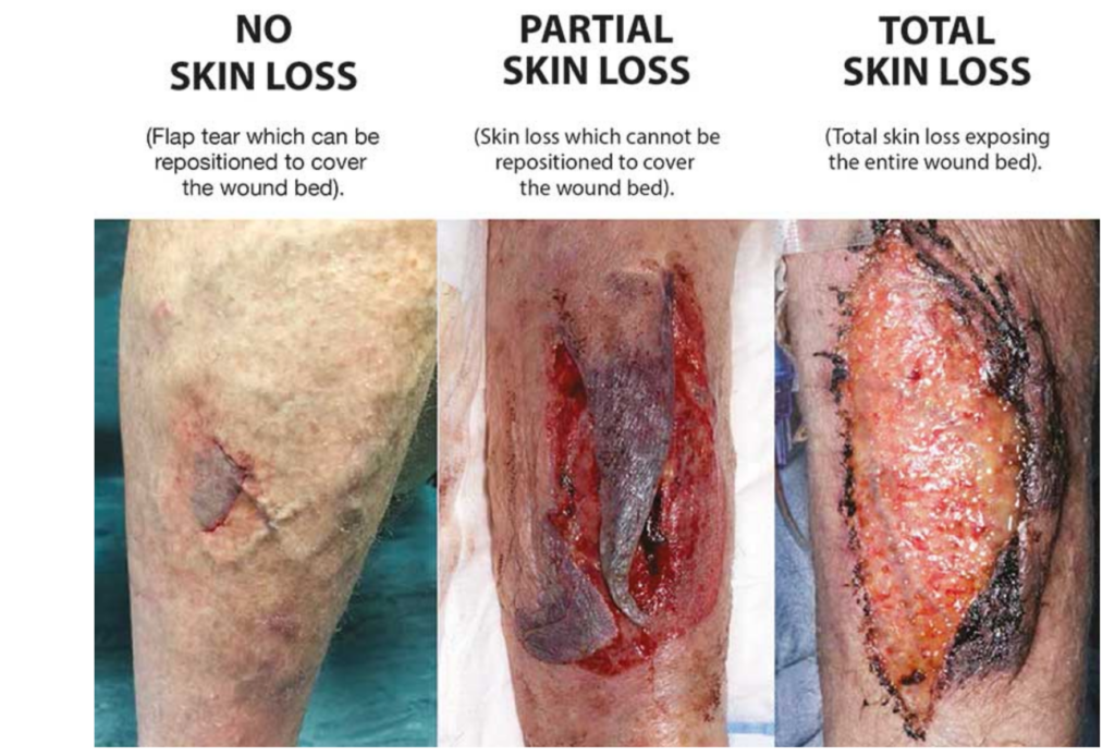
(https://richardsonhealthcare.com/istap-classification-skin-tears/)
General Management:
- Goals:
-
- Protect the affected skin from further injury and possible infections.
- Maintain a moist wound environment to promote healing.
- Cleansing:
- Rinse wounds thoroughly with tap water or normal saline.
- Heavily contaminated wounds should be irrigated at ≥7 psi, which can be achieved using a 10-50 mL syringe with a splatter shield.
- Wound Approximation:
- The skin tear flap should be repositioned as closely as possible to its original position.
- Excess blood and serum should be expressed (milked) by applying gentle pressure before attempting closure.
- Tetanus Prophylaxis:
- Ensure the patient’s tetanus immunization status is up to date.
- Prophylactic Antibiotics:
- While guidelines on prophylactic antibiotic use for skin tears are limited, antibiotics may be reasonable for:
- Heavily contaminated wounds.
- Wounds with a delay in closure.
- Patients with comorbidities that increase infection risk.
- While guidelines on prophylactic antibiotic use for skin tears are limited, antibiotics may be reasonable for:
Anesthesia:
- Challenges
- Large surface area may make typical infiltrative local anesthesia techniques impractical.
- Skin flaps, especially those with abnormal color, may have a theoretical risk of increased risk of tissue ischemia and poor wound healing with the use of large amounts of epinephrine-containing local anesthetics.
- Topical Anesthesia
- Benefits:
- Application is quick and relatively painless.
- Provides effective pain relief over large surface areas.
- Hydrates the skin.
- Options:
- 2% Topical Lidocaine Jelly (Commonly carried as Uro-Jet) contains hydroxypropylmethylcellulose, a water-soluble solution that retains water and acts as an emulsifier.
- Safety Considerations:
- Topical Lidocaine can lead to systemic absorption and toxicity.
- Maximum recommended dose of Lidocaine is 4.5 mg/kg (15 mL of 2% Lidocaine Jelly in the average 70 kg adult).
- Benefits:
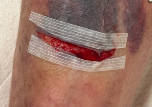
Category 1 Skin Tears (Minimal or No Tissue Loss):
- Goal: Primary Closure
- Closure Methods:
- Surgical tape (Steri-Strips®)
- Cyanoacrylate-based topical adhesives (contraindicated if the wound extends beyond the dermis).
- Sutures, if necessary (for high-tension areas like joints).
- Dressing:
- Use a non-adherent dressing (coat with topical antimicrobial) held in place with wrapped dry-gauze or tubular net.
- Mark the recommended direction of dressing removal to prevent further skin trauma.
Surgical Tape vs. Sutures
- Surgical tape or adhesive are preferred, as they are less painful, quicker to apply, and cause less tissue trauma.
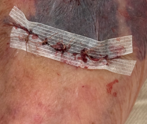
- Suturing of skin tears can possess a unique challenge, as the increased skin frailty associated with skin tears often results in a “cheese-wire” effect where the suture tears through the skin.
- Wounds in areas of high tension (ie., retracted skin, tissue loss, large joints, etc.) may benefit from sutures to reduce the risk of wound dehiscence and obtain optimal wound alignment.
- One technique that can help to prevent suture “cheese-wiring” (where sutures tear fragile skin), is to place Steri-Strips® horizontally along wound margins before suturing through them.
Category 2 & 3 Skin Tears (Partial or Complete Tissue Loss):
- Category 2 skin tears with minimal tissue loss and reasonable approximation may be managed as described for Category 1 skin tears.
- Category 2 (Significant Tissue Loss) and Category 3:
- Goal: Secondary Closure (Conservative management with dressings).
- The goal is to create a moist, artificial barrier to prevent infection and promote generation of granulation tissue.
- Wound Protection:
- Apply a topical antimicrobial.
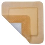
- Use an absorbent dressing, such as:
- Hydrocolloids
- Xeroform® (Nylon-impregnated gauze)
- Silicone-coated foam dressings
- Hydrocolloids
- Goal: Secondary Closure (Conservative management with dressings).

-
- Dressing Protocol:
- Keep the primary dressing in place for 5-7 days.
- Cover with a secondary absorbent gauze dressing, changed daily as needed.
- Secure with dry gauze or a tubular net for support.
- Alternative Approach:
- Apply cyanoacrylate-based topical adhesive directly to the wound bed to form an in situ dressing.
- Dressing Protocol:
- Consultation and Referral
- All category 2 and 3 skin tears should have specialist referral as they are at high risk of failing conservative measures and often involve eventual surgical debridement and grafting.
- When available, early consultation or referral to wound care surgery from the ED can lead to improved outcomes for patients including reduced inpatient stays and associated wound complications.
Disposition:
- When determining disposition, take into consideration factors that led to the injury:
- Fall due to a medical etiology (ex., syncope).
- Patterns of multiple, frequent falls may be unsafe to discharge home.
- Concern for elder abuse or neglect.
- In general, most wounds that are managed in the ED can be discharged home, however, patients with large surface area wounds may benefit from inpatient admission.
- Consider admission in patients who have new functional limitations due to injury.
Prevention Strategies:
- Educate patients and families on prevention strategies to prevent future skin tears.
- Optimize environmental safety (e.g., remove fall hazards, improve lighting).
- Padding rails, wheelchair arms, and leg supports.
- Proper transfer techniques.
- Use protective garments (e.g., long sleeves, shin guards for high-risk patients).
References:
- Bragg, K., & Fox, H. (2019, December 11). Trick of Trade: Topical lidocaine jelly takes the tears out of skin tears and road rash. ALiEM. Retrieved from https://www.aliem.com/trick-of-trade-topical-lidocaine-jelly-skin-tears-road-rash/
- Singer, A. J., & Dagum, A. B. (2008). Current management of acute cutaneous wounds. The New England Journal of Medicine, 359(10), 1037–1046. https://doi.org/10.1056/NEJMra0707253
- Vandervord, J. G., Tolerton, S. K., Campbell, P. A., Darke, J. M., & Loch-Wilkinson, A.-M. V. (2016). Acute management of skin tears: A change in practice pilot study. International Wound Journal, 13(1), 59–64. https://doi.org/10.1111/iwj.12227
- Xu, X., Lau, K., Taira, B. R., & Singer, A. J. (2009). The current management of skin tears. The American Journal of Emergency Medicine, 27(6), 729–733. https://doi.org/10.1016/j.ajem.2008.05.015
