- Author: Mary Weber
- Editor: Jonathan Kobles, MD
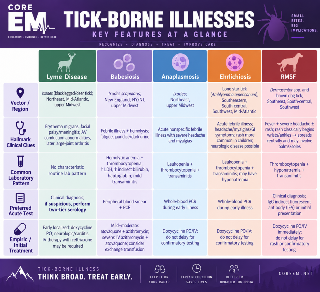
Lyme Disease
Rapid Reference
- Primary Manifestations: Early localized disease is characterized by erythema migrans (EM); disseminated or late disease may manifest with multiple EM lesions, facial nerve palsy, meningitis or radiculoneuropathy, carditis with AV conduction abnormalities, and arthritis.
- Clinical Pattern: EM is a clinical diagnosis and should be treated without awaiting serologic confirmation. Serologic testing may be negative early in infection; suspected disseminated or late disease should undergo two-tier serologic testing. In Lyme carditis, ECG findings range from PR prolongation to second- or third-degree AV block and may fluctuate rapidly.
- Treatment of Early Localized Disease: Doxycycline 100 mg PO twice daily for 10 days; amoxicillin or cefuroxime are alternatives.
- Treatment of Neurologic Lyme Disease: Lyme meningitis, cranial neuropathy, and radiculoneuropathy may be treated with oral doxycycline or IV ceftriaxone for 14-21 days, with route and disposition determined by severity. Parenchymal brain or spinal cord involvement warrants IV therapy.
- Treatment of Lyme Carditis: Severe carditis requires hospitalization with continuous cardiac monitoring and IV ceftriaxone, with transition to oral therapy after clinical improvement; temporary pacing may be required for symptomatic bradycardia or high-grade AV block.
- Treatment of Lyme Arthritis: Doxycycline, amoxicillin, or cefuroxime PO for 28 days.
Introduction
- Lyme disease is the most common vector-borne disease in the United States and is caused by Borrelia burgdorferi, a spirochete transmitted by infected Ixodes ticks.
- Clinical manifestations vary according to the stage of infection and range from the characteristic erythema migrans (EM) rash to neurologic, cardiac, and rheumatologic disease.
- Lyme disease is a clinical diagnosis; serologic testing may initially be negative, and prompt antibiotic therapy is indicated to prevent complications.
Epidemiology
Geographic Distribution
- Lyme disease is concentrated primarily in the Northeast, Mid-Atlantic, and upper Midwest regions of the United States.
- Historically, approximately 90% of reported infections have been acquired in a relatively small number of endemic states in these regions.
- Cases occurring outside endemic regions are frequently associated with travel to an endemic area.
- Exposure to wooded, brushy, and grassy environments, including residential yards adjacent to these environments, increases the risk of tick exposure.
Disease Burden
- In 2023, 89,470 confirmed and probable Lyme disease cases were reported to CDC.
- Surveillance data underestimate the total number of patients diagnosed and treated for Lyme disease.
- The CDC estimates that approximately 476,000 people are diagnosed and treated for Lyme disease annually in the United States.
Pathophysiology
Ticks can acquire B. burgdorferi while feeding on infected reservoir hosts, particularly small mammals such as the white-footed mouse.
-
- During feeding, B. burgdorferi migrates from the tick midgut to the salivary glands and is transmitted into the host. The organism initially spreads through the skin and may subsequently disseminate, producing neurologic, cardiac, musculoskeletal, and constitutional manifestations.
- The probability of transmission increases with attachment duration and is low during the first 24 hours; most transmission occurs after approximately 36–48 hours of attachment.
Clinical Manifestations
Lyme disease has traditionally been divided into three clinical stages, which may overlap.
Early Localized
Erythema migrans (EM) is the hallmark manifestation of early Lyme disease. EM typically develops 3–30 days after the tick bite, with onset commonly occurring approximately 1–2 weeks after exposure.
The lesion typically:
- Begins as a red macule or papule near the site of the tick bite
- Expands gradually and is typically ≥5 cm; lesions may become very large.
- May be uniformly erythematous or demonstrate central clearing
- Can occasionally have vesicular or necrotic central changes
- Is usually minimally symptomatic, although mild pruritus, burning, or discomfort may occur
Patients may also experience:
- Fever
- Malaise
- Fatigue
- Headache
- Myalgias
- Arthralgias
- Regional lymphadenopathy
- Neck discomfort or stiffness
Early Disseminated Disease
The most common manifestation of early disseminated disease is the development of multiple erythema migrans, secondary to hematogenous dissemination.
Neurologic manifestations may include:
- Cranial neuropathy (most commonly facial nerve palsy)
- Lymphocytic meningitis (headache and neck stiffness)
- Radiculoneuritis
- Papilledema and Elevated Intracranial Pressure (ICP)
Cardiac Manifestations
Atrioventricular conduction disease may develop, in which patients present with:
- Lightheadedness
- Palpitations
- Syncope
- Dyspnea
- Chest discomfort
ECG findings range from PR prolongation to second- or third-degree AV block and may fluctuate rapidly.
Late Lyme Disease
Lyme arthritis is one of the most characteristic manifestations of late disease.
Typical findings include:
- Objective joint swelling
- Monoarticular or oligoarticular involvement
- Predominantly large joints
- The knee is particularly common
- Joint effusion may be substantial
Additionally, disseminated neurologic disease may present as encephalomyelitis and polyneuropathy.
Laboratory Studies
Early localized Lyme disease (erythema migrans) is primarily a clinical diagnosis and does not require serologic confirmation prior to initiating treatment. High clinical suspicion should be for those with:
- Epidemiologically plausible tick exposure and
- A skin lesion clinically compatible with erythema migrans
Serologic Testing
- CDC recommends a two-tier serologic testing algorithm using FDA-cleared assays. Standard two-tier testing uses an enzyme immunoassay (EIA) followed, when indicated, by an immunoblot; modified two-tier testing uses two sequential EIAs.
- Antibody tests may be falsely negative during the first several weeks of infection. Once antibodies develop, they may remain detectable for months to years and should not be used to document cure.
Treatment
Early Localized
- Treat uncomplicated EM with one of the following oral regimens:
- Doxycycline: 100 mg PO BID for 10 days
- Amoxicillin: 500 mg PO TID for 14 days
- Cefuroxime: 500 mg PO BID for 14 days
- Azithromycin is generally considered a second-line option when patients cannot take doxycycline or beta-lactam antibiotics.
Neurologic Lyme Disease
- Lyme meningitis, cranial neuropathy, and radiculoneuropathy may be treated with oral doxycycline or IV ceftriaxone, cefotaxime, or penicillin G for 14–21 days.
- Route should be individualized based on the specific manifestation, severity, tolerability, and adherence; parenchymal brain or spinal cord involvement warrants IV therapy.
Lyme Carditis
- Hospitalized patients with significant Lyme carditis should be started on IV ceftriaxone, with transition to oral therapy after clinical improvement. Total antibiotic duration is generally 14–21 days.
- Patients with symptomatic bradycardia requiring pacing should generally receive temporary pacing rather than immediate permanent pacemaker placement, as conduction abnormalities typically improve with treatment.
Lyme Arthritis
- Initial Lyme arthritis is generally treated with 28 days of oral antibiotic therapy. Agents include
- Doxycycline
- Amoxicillin
- Cefuroxime axetil
Post-Exposure Prophylaxis
Consider prophylaxis for a high-risk Ixodes bite when::
- The exposure occurred in a highly endemic area.
- The tick was attached for ≥36 hours.
- Doxycycline can be started within 72 hours of tick removal.
- Doxycycline: single dose
- Adults: 200 mg PO once
- Children: 4.4 mg/kg PO once, maximum 200 mg
Side Effect of Treatment: Jarisch-Herxheimer Reaction
Patients beginning antibiotic therapy for Lyme disease may develop a transient inflammatory response to treatment associated with spirochetal killing. Patients should be counseled that this is not an allergic or medication reaction.
Symptoms can include:
- Fever
- Chills
- Sweating
- Headache
- Myalgias
- Transient worsening of existing symptoms
Symptoms generally occur shortly after beginning therapy and are self-limited. Supportive treatment, including antipyretics and NSAIDs when appropriate, can be used.
Disposition
- Patients with early localized disease may be discharged with outpatient antibiotic therapy and routine primary-care follow-up.
- Those with suspected Lyme carditis require hospitalization for continuous cardiac monitoring, and potentially ICU-level care if they become hemodynamically unstable, requiring pacing.
- Patients with neurologic Lyme disease require admission for IV antibiotic therapy and monitoring.
Prognosis
- The prognosis for appropriately treated Lyme disease is generally excellent, particularly when therapy is initiated during early disease.
- Most patients treated during early infection recover rapidly and completely.
- Some patients experience prolonged nonspecific symptoms such as:
- Fatigue
- Musculoskeletal pain
- Cognitive complaints
Babesiosis
Rapid Reference
- Primary Manifestations: Nonspecific febrile illness with fever, chills, headache, malaise, and myalgias; severe disease may progress to hemolytic anemia, ARDS, renal or hepatic dysfunction, shock, and multiorgan failure.
- Clinical Pattern: Laboratory studies commonly demonstrate hemolytic anemia, thrombocytopenia, elevated indirect bilirubin and LDH, reduced haptoglobin, and mild transaminitis; renal dysfunction may occur in severe disease. Diagnosis is confirmed by peripheral blood smear demonstrating intraerythrocytic parasites, including ring forms or the characteristic Maltese cross, or by Babesia PCR.
- Treatment of Mild-to-Moderate Disease: Atovaquone 750 mg PO twice daily + azithromycin for 7-10 days.
- Treatment of Severe Disease: Hospitalization with IV azithromycin + oral atovaquone; IV clindamycin + oral quinine is an alternative. Consider exchange transfusion for high-grade parasitemia (>10%), severe hemolytic anemia, or severe pulmonary, renal, or hepatic compromise.
Introduction
- Babesiosis is a tick-borne infection caused by intraerythrocytic protozoan parasites of the genus Babesia.
- In the United States, Babesia microti is the most common species causing human disease and is maintained in nature primarily through small-mammal reservoirs, including the white-footed mouse.
- Babesia parasites invade and replicate within red blood cells, and, therefore, produce a clinical syndrome that can resemble malaria, with fever, hemolytic anemia, and parasitemia.
- Transmission occurs primarily through the bite of an infected Ixodes scapularis (blacklegged or deer tick), or rarely through blood transfusion, organ transplantation, or vertical transmission.
Epidemiology
- In the United States, babesiosis occurs predominantly in:
- New England
- New York and New Jersey
- Upper Midwest, particularly Wisconsin and Minnesota
- In 2022, reported incidence was highest in Maine, Rhode Island, and Vermont.
- Most tick-borne cases occur during the late spring through early fall, with a peak during the summer months.
- Reported babesiosis cases have increased substantially over time.
Pathophysiology
During a blood meal, an infected tick introduces Babesia sporozoites into the human host.
The parasites then:
- An infected tick introduces Babesia sporozoites into the human host, where the parasites then enter circulating erythrocytes, undergo asexual replication, and rupture the infected cell.
- The combination of erythrocyte invasion and destruction produces the characteristic hemolytic anemia associated with babesiosis.
Clinical Manifestations
Babesiosis ranges from completely asymptomatic infection to fulminant, life-threatening disease. The incubation period is generally 1-4 weeks following a tick bite, and 3 weeks to 6 months after transfusion of contaminated blood products.
Many infections are asymptomatic, particularly in younger, immunocompetent patients.
Mild-to-moderate disease typically presents as a nonspecific febrile illness. Symptoms include:
- Fever
- Chills
- Sweats
- Fatigue
- Malaise
- Headache
- Myalgias
- Arthralgias
- Anorexia
Additional symptoms may include:
- Nausea
- Vomiting
- Abdominal discomfort
- Dry cough
- Dyspnea
- Dark urine
- Weight loss
Physical examination may demonstrate:
- Splenomegaly
- Hepatomegaly
- Jaundice
Potential complications from severe disease include:
- Severe hemolytic anemia
- Acute respiratory distress syndrome
- Pulmonary edema
- Acute kidney injury or renal failure
- Hepatic dysfunction
- Disseminated intravascular coagulation
- Shock
- Congestive heart failure
- Splenic infarction
- Splenic rupture
- Altered mental status
- Multiorgan failure
- Death
Laboratory Studies
- A diagnosis should be suspected from the combination of:
- Compatible clinical illness
- Epidemiologic exposure
- Hemolytic anemia
- Thrombocytopenia
- Definitive diagnosis generally requires direct detection of the organism with peripheral blood smear or PCR.
- Microscopic examination of Giemsa- or Wright-stained thin blood smears may demonstrate intraerythrocytic parasites. Typical findings of smear include:
- Findings include pleomorphic intraerythrocytic ring forms and, less commonly, tetrads (‘Maltese crosses’). Parasitemia may be low; repeat smears or PCR may be necessary when suspicion remains high.
- Serologic testing can support the diagnosis but cannot distinguish active from previous infection.
Additional Laboratory Studies
- CBC with differential
- Peripheral smear
- Reticulocyte count
- CMP
- Total and direct bilirubin
- LDH
- Haptoglobin
- Urinalysis
- Babesia PCR
- Coagulation studies in severe disease
- Testing for other tick-borne illness
Common laboratory findings include:
- Hemolytic anemia
- Thrombocytopenia
- Normal or mildly decreased leukocyte count
- Elevated reticulocyte count
- Elevated LDH
- Elevated indirect bilirubin
- Reduced haptoglobin
- Mild-to-moderate transaminitis
- Elevated BUN
- Elevated creatinine
- Hemoglobinuria
- Hematuria
- Increased urobilinogen
Treatment
-
- Mild to Moderate Babesiosis: 7-10 days treatment
- Immunocompetent patients with mild-to-moderate babesiosis: atovaquone 750 mg PO twice daily plus azithromycin 500 mg PO on day 1, then 250 mg daily, typically for 7–10 days.
- Alternative regimens include Clindamycin + Quinine
- Immunocompetent patients with mild-to-moderate babesiosis: atovaquone 750 mg PO twice daily plus azithromycin 500 mg PO on day 1, then 250 mg daily, typically for 7–10 days.
- Mild to Moderate Babesiosis: 7-10 days treatment
- Severe Babesiosis
-
- IV azithromycin + oral atovaquone or alternatively IV clindamycin + oral quinine
- Exchange Transfusion
- High-grade parasitemia >10%
and/or - Severe hemolytic anemia
and/or - Severe pulmonary compromise
- Severe renal compromise
- Severe hepatic compromise
- High-grade parasitemia >10%
Disposition
- Patients who can tolerate oral therapy with evidence of mild disease may be discharged with outpatient therapy and close follow-up.
- Those who are immunocompromised or have evidence of hemolysis, renal, hepatic, or cardiovascular complications should be admitted for IV therapy.
- Patients who develop ARDS, shock, or organ dysfunction require ICU-level care and potential exchange transfusion.
Prognosis
- The prognosis is excellent for otherwise healthy patients with mild-to-moderate babesiosis who receive appropriate treatment.
- Symptoms usually improve within several days of initiating therapy.
- Severe and fatal disease occurs disproportionately among:
- Asplenic patients
- Older adults
- Highly immunocompromised patients
- Patients with major underlying comorbidities
References
- Waked R, Krause PJ. Human Babesiosis. Infect Dis Clin North Am. 2022;36(3):655-670. doi:10.1016/j.idc.2022.02.009.
- Krause PJ. Human babesiosis. Int J Parasitol. 2019;49(2):165-174. doi:10.1016/j.ijpara.2018.11.007.
- Krause PJ, Auwaerter PG, Bannuru RR, et al. Clinical Practice Guidelines by the Infectious Diseases Society of America: 2020 Guideline on Diagnosis and Management of Babesiosis. Clin Infect Dis. 2021.
- Swanson M, Pickrel A, Williamson J, Montgomery S. Trends in reported babesiosis cases—United States, 2011–2019. MMWR Morb Mortal Wkly Rep. 2023;72:273-277.
- Moritz ED, Winton CS, Tonnetti L, et al. Screening for Babesia microti in the U.S. blood supply. N Engl J Med.2016;375(23):2236-2245.
- Allred DR. Babesiosis: persistence in the face of adversity. Trends Parasitol. 2003;19(2):51-55.
- Centers for Disease Control and Prevention. Babesiosis: Clinical Overview; Clinical Care; Data and Statistics; DPDx—Babesiosis.
- U.S. Food and Drug Administration. Transfusion/Donation Fatalities.
Anaplasmosis
Rapid Reference
- Primary Manifestations: Nonspecific acute febrile illness with fever, headache, myalgias, and malaise; severe disease may progress to coagulopathy, AKI, respiratory failure, and multiorgan failure.
- Clinical Pattern: Leukopenia, thrombocytopenia, and elevated hepatic transaminases are characteristic laboratory findings. Whole-blood PCR is the preferred confirmatory test during early illness; peripheral smear may demonstrate morulae within granulocytes. Treatment should not be delayed while awaiting confirmatory testing.
- Treatment of Mild Disease: Doxycycline 100 mg PO every 12 hours for 10-14 days and for at least 3 days after defervescence.
- Treatment of Severe Disease: Hospitalization with doxycycline 100 mg IV every 12 hours, with transition to oral therapy following clinical improvement; treat for 10-14 days and for at least 3 days after defervescence.
Introduction
- Anaplasmosis, also known as human granulocytic anaplasmosis (HGA), is a tick-borne illness caused by Anaplasma phagocytophilum, an obligate intracellular bacterium transmitted primarily through the bite of infected Ixodes ticks.
- Anaplasmosis frequently presents as a nonspecific acute febrile illness and can initially be mistaken for a viral syndrome.
- Recognition in the emergency department is important because delayed antimicrobial therapy increases the risk of severe disease, while appropriate treatment often produces rapid clinical improvement.
Epidemiology
- In the United States, anaplasmosis occurs predominantly in the:
- Northeast
- Upper Midwest
- Most reported U.S. cases occur in the Northeast and upper Midwest
- High endemic states include:
- Maine
- Massachusetts
- Minnesota
- New Jersey
- New York
- Pennsylvania
- Vermont
- Wisconsin
- The geographic distribution overlaps substantially with that of Lyme disease because both infections can be transmitted by Ixodes ticks. Rate of coinfection ranges between 2% and 11.7%.
- The incidence of anaplasmosis has increased substantially since national surveillance began. While only hundreds of cases were reported annually during the early 2000s, in 2023, 7280 cases were reported to the CDC.
Pathophysiology
- Ticks, primarily Ixodes scapularis and Ixodes pacificus, acquire A. phagocytophilum by feeding on an infected host; A. phagocytophilum is transmitted to humans through tick saliva.
- The bacterium infects circulating neutrophils and other granulocytes and replicates within the morulae.
- Tick attachment for 24 hours is needed to transmit disease to humans.
Clinical Manifestations
Symptoms typically begin approximately 5–14 days after an infected tick bite. Many patients do not recall a tick bite.
The clinical spectrum ranges from asymptomatic infection or mild illness to severe multisystem disease.
Early Illness
The most common presentation is a nonspecific acute febrile illness. Symptoms include:
- Fever
- Chills
- Rigors
- Severe headache
- Malaise
- Fatigue
- Myalgias
- Arthralgias
- Nausea
- Vomiting
- Diarrhea
- Anorexia
Less frequently reported findings include:
- Cough
- Neck stiffness
- Confusion
- Elevated creatinine
- Anemia
- Neurologic manifestations
Severe Disease
Delayed treatment can lead to severe multisystem disease, with reported complications including:
- Respiratory failure
- Acute kidney injury
- Disseminated intravascular coagulation-like coagulopathy
- Hemorrhage
- Rhabdomyolysis
- Peripheral neuropathy
- Meningoencephalitis
- Multiorgan failure
- Death
Laboratory Studies
- The ED diagnosis is often clinical. Anaplasmosis should be suspected in patients with:
- Acute febrile illness
- Compatible epidemiologic exposure
- Residence in or travel to an endemic region
- Recent outdoor or tick exposure
- PCR performed on whole blood is the preferred confirmatory test during early illness.
- Is most sensitive during the first week of illness
- Provides evidence of active infection when positive
- Becomes less sensitive shortly after appropriate antibiotic therapy begins
- A negative PCR does not exclude anaplasmosis when clinical suspicion remains high.
- A Wright- or Giemsa-stained peripheral blood smear may demonstrate characteristic morulae.
- Indirect Fluorescent Antibody (IFA) for A. phagocytophilum IgG is the most sensitive confirmatory test but acute serology is often negative during the first week and should not delay treatment.
- A fourfold rise in IgG antibody titer provides strong evidence of recent infection.
Additional Laboratory Evaluation
- CBC with differential
- Leukopenia
- Thrombocytopenia
- Mild Anemia
- Comprehensive metabolic panel
- Elevated Creatinine
- Hepatic transaminases
- Elevated AST/ALT
Treatment
- Doxycycline is the treatment of choice for adults and children of all ages. Empiric therapy should be initiated when anaplasmosis is clinically suspected.
- Adults and Children ≥45 kg: 100 mg PO/IV every 12 hours
- Children <45 kg: 2.2 mg/kg PO/IV every 12 hours, maximum 100 mg per dose
- Patients with suspected anaplasmosis are generally treated for 10–14 days to provide an appropriate treatment duration for possible concurrent Borrelia infection.
- Treatment should continue for at least 3 days after defervescence.
- Rifampin may be considered for mild anaplasmosis in selected patients with a true contraindication to doxycycline, including some pregnant patients, after infectious-disease consultation. It does not treat RMSF or Lyme disease, so alternative diagnoses and coinfection must be excluded.
Disposition
- Those who are hemodynamically stable with mild illness may be discharged with oral antibiotic therapy and a recommendation for primary-care follow-up.
- ICU-level care is indicated for those with severe complications, such as respiratory failure, severe coagulopathy, shock, or multiorgan failure.
Prognosis:
- The prognosis of appropriately treated anaplasmosis is generally excellent.
- Overall case-fatality rate is less than 1%, although severe and fatal disease can occur.
- Most patients demonstrate significant clinical improvement within 24–48 hours of starting doxycycline.
Ehrlichiosis
Rapid Reference
- Primary Manifestations: Nonspecific acute febrile illness with fever, headache, myalgias, and malaise; a maculopapular or petechial rash may occur, particularly in children. Severe disease may progress to CNS involvement, AKI, respiratory failure, shock, HLH, or multiorgan failure.
- Clinical Pattern: Leukopenia, thrombocytopenia, elevated hepatic transaminases, and hyponatremia are characteristic laboratory findings. Whole-blood PCR is preferred during early illness; peripheral smear may demonstrate morulae within monocytes. Treatment should not be delayed while awaiting confirmatory testing.
- Treatment of Mild Disease: Doxycycline 100 mg PO every 12 hours for at least 5-7 days and until afebrile for ≥72 hours with clinical improvement.
- Treatment of Severe Disease: Hospitalization with doxycycline 100 mg IV every 12 hours and supportive care; transition to oral therapy following clinical improvement. Continue treatment for at least 5-7 days and until afebrile for ≥72 hours with clinical improvement.
Introduction
- Ehrlichiosis is a tick-borne infection caused by obligate intracellular gram-negative bacteria of the genus Ehrlichia.
- In the United States, human ehrlichiosis is primarily caused by:
- Ehrlichia chaffeensis
- Ehrlichia ewingii
- Ehrlichia muris eauclairensis
- E. chaffeensis is the most common cause of human monocytic ehrlichiosis (HME) and preferentially infects monocytes and macrophages.
- The infection is transmitted primarily by the lone star tick (Amblyomma americanum), although some erichlia species can be transmitted by the Ixodes tick
Epidemiology
Geographic Distribution
- Ehrlichiosis occurs predominantly in the South-central United States, Southeastern United States, and Mid-Atlantic region
- Reported E. chaffeensis ehrlichiosis is concentrated in south-central, southeastern, and mid-Atlantic states, broadly following the range of the lone star tick.
- Ehrlichia muris eauclairensis is transmitted by the blacklegged tick (Ixodes scapularis), with most recognized U.S. cases occurring in Minnesota and Wisconsin.
- Rare transmission has been reported through blood transfusion and organ transplantation.
Pathophysiology
- After transmission through a tick bite, Ehrlichia organisms enter host leukocytes through endocytosis and form morulae.
- The bacteria alter normal host-cell signaling and immune responses, which promote a systemic inflammatory response.
- E. chaffeensis primarily targets monocytes and macrophages.
Clinical Manifestations
Symptoms generally develop 5–14 days after an infected tick bite, although most patients do not remember a history of tick exposure.
The clinical spectrum ranges from asymptomatic or mild infection to rapidly progressive multisystem disease.
Early symptoms are generally nonspecific, and may include:
- Fever
- Chills or rigors
- Headache
- Malaise
- Fatigue
- Myalgias
- Arthralgias
- Nausea
- Vomiting
- Diarrhea
- Anorexia
- Abdominal discomfort
Rash when present may be an important distinguishing feature of ehrlichiosis. Unlike erythema migrans, the rash of ehrlichiosis is not caused by direct bacterial infection of the skin and is believed to reflect the systemic inflammatory response.
It develops in up to approximately 60% of children and less than 30% of adults
The rash generally appears around the fifth day of illness, so its absence early in the clinical course does not exclude disease.
Morphology may include:
- Maculopapular eruption
- Petechiae
- Diffuse erythema
The rash may involve the trunk or extremities and can include the palms and soles; morphology and distribution are variable.
Severe neurologic involvement can occur, particularly in severe E. chaffeensis infection, and should increase concern for disseminated disease and prompt consideration of hospitalization.
Manifestations may include:
- Severe headache
- Confusion
- Meningismus
- Encephalopathy
- Seizures
- Meningitis or meningoencephalitis
Untreated ehrlichiosis can progress rapidly. Potential complications include:
- Sepsis-like syndrome
- Septic shock
- Acute respiratory distress syndrome
- Respiratory failure
- Acute kidney injury
- Hepatic dysfunction
- Coagulopathy
- Disseminated intravascular coagulation
- Meningoencephalitis
- Hemophagocytic lymphohistiocytosis (HLH)
- Multiorgan failure
- Death
Risk of severe disease increases substantially when doxycycline is delayed. Additionally, severe or fatal infection is more likely in patients with:
- Delayed initiation of doxycycline
- Older age
- Immunosuppression
- Malignancy
- Solid-organ transplantation
- Significant chronic illness
- Very young age
Laboratory Studies
- Ehrlichiosis is initially a clinical diagnosis.
- Whole-blood PCR: the preferred confirmatory diagnostic test during early illness. PCR is:
- Most sensitive during the first week of illness
- Most useful before or shortly after antibiotics are started
- Evidence of active infection when positive
- Serology: Obtained via IgG indirect fluorescent antibody (IFA), it consists of an acute specimen obtained during the first 2 weeks of illness and a convalescent specimen obtained several weeks later.
- A fourfold rise in IgG titer between acute and convalescent samples provides strong evidence of recent infection.
- A peripheral smear may reveal morulae within monocytes in E. chaffeensis infection, but sensitivity is insufficient to rule out ehrlichiosis when morulae are absent.
Additional ED Laboratory Evaluation
- CBC with differential
- Leukopenia
- Thrombocytopenia
- Mild anemia
- Comprehensive metabolic panel
- Hyponatremia
- Elevated Creatinine
- Liver Function Tests
- Elevated AST/ALT
- CSF from Lumbar Puncture
- Mildly Elevated Protein
- Mild Lymphocytic or Monocytic Pleocytosis
For severely ill patients, also consider:
- Coagulation studies
- Lactate
- Blood cultures
- LDH
- Ferritin if HLH is suspected
- Additional sepsis evaluation as clinically indicated
Treatment
- Doxycycline is the first-line treatment for adults and children of all ages.
- Adults: 100 mg PO/IV q12 hours
- Children <45kg: 2.2 mg/kg PO/IV q12 hours, maximum 100 mg per dose
- Delay in appropriate treatment poses a substantially greater risk than the theoretical dental concerns associated with older tetracycline drugs.
- Treatment should last 7-10 days, and continue until the patient has remained afebrile for at least 72 hours with clinical improvement.
- Do not delay doxycycline while awaiting PCR or serology.
- Fever usually resolves within 24–48 hours after starting doxycycline. Failure to improve should prompt reconsideration of diagnosis or potential for coinfection.
Disposition
- Stable patients who can tolerate oral therapy may be discharged with outpatient therapy and primary-care follow-up.
- Patients with neurologic involvement, severe cytopenias, inability to tolerate oral therapy, or history of immunocompromise require admission for IV therapy.
- Patients experiencing DIC, ARDS, severe CNS disease or multiorgan failure require ICU admission.
Rocky Mountain Spotted Fever
Rapid Reference
- Primary Manifestations: Acute febrile illness with severe headache, myalgias, malaise, and GI symptoms; a characteristic rash may develop on the wrists and ankles and spread centrally, often involving the palms and soles. Rash may be absent early or throughout the illness. Severe disease may progress to encephalitis, ARDS, shock, and multiorgan failure.
- Clinical Pattern: Thrombocytopenia, hyponatremia, and elevated hepatic transaminases may occur, although laboratory abnormalities may be absent early. Serology is frequently negative during the first week; treatment should not be delayed while awaiting confirmatory testing.
- Treatment of Mild Disease: Doxycycline 100 mg PO every 12 hours for at least 5-7 days and for ≥3 days after defervescence and clinical improvement.
- Treatment of Severe Disease: Hospitalization with doxycycline 100 mg IV every 12 hours and supportive care; transition to oral therapy following clinical improvement. Do not delay doxycycline while awaiting diagnostic confirmation.
Introduction
- Rocky Mountain spotted fever (RMSF) is a potentially life-threatening tick-borne infection caused by Rickettsia rickettsii, an obligate intracellular gram-negative coccobacillus.
- RMSF is the most severe spotted fever rickettsiosis in the United States and can progress rapidly from a nonspecific febrile illness to multiorgan failure and death.
- The organism primarily infects vascular endothelial cells, producing widespread vasculitis, increased vascular permeability, edema, and tissue injury.
- Treatment must be started immediately when there is a high clinical suspicion for RMSF
- Untreated RMSF historically had case-fatality rates of approximately 20%–30%
- Delayed doxycycline, particularly beyond day 5 of illness, markedly increases the risk of death.
Epidemiology
- RMSF occurs throughout much of the United States but has historically been most strongly associated with the South-central and Southeastern United States, despite its name.
- More than half of reported spotted fever rickettsiosis cases arise from North Carolina, Oklahoma, Arkansas, Tennessee, and Missouri.
- Tick species responsible for transmitting rickettsial infections include:
- American Dog Tick (eastern and central USA)
- Rocky Mountain Wood Tick (western USA)
- Brown Dog Tick (southwestern USA)
Pathophysiology
- After inoculation through the tick bite, R. rickettsii disseminates hematogenously and preferentially infects and replicates within vascular endothelial cells. This produces endothelial injury, vascular inflammation, and increased capillary permeability.
- Generally, there is a 2–14–day incubation period. A shorter interval between exposure and symptom onset is associated with more severe disease.
Clinical Manifestations
Early disease may present with nonspecific symptoms, including:
- Fever
- Severe headache
- Malaise
- Myalgias
- Chills
- Nausea
- Vomiting
- Abdominal pain
- Anorexia
Additional findings may include:
- Nonproductive cough
- Conjunctival injection
- Photophobia
- Restlessness
- Peripheral edema
The characteristic rash typically develops 2-5 days after the fever begins and is progressive. Fewer than half of patients have a rash during the first 3 days of illness, and a minority never develop one; absence of rash must not delay treatment.
When present, the rash may appear as:
- Small, blanching, pink macules on the wrists, ankles, and forearms
- Progresses to spread centrally and to include the palms and soles
- Facial rash is uncommon
- Petechiae present later in the course of illness and signify vascular injury.
Neurologic involvement, another marker of more severe disease, can closely resemble bacterial/viral meningoencephalitis, and present as:
- Severe headache
- Meningismus
- Confusion
- Encephalopathy
- Seizures
- Focal neurologic deficits
- Coma
Severe or untreated disease can rapidly progress to:
- Encephalitis
- ARDS
- Noncardiogenic pulmonary edema
- Myocarditis
- Arrhythmias
- Acute kidney injury
- Hepatic dysfunction
- DIC
- Shock
- Peripheral ischemia
- Tissue necrosis
- Gangrene
- Multiorgan failure
- Death
Risk factors for the development of severe disease include:
- Delayed doxycycline treatment
- Children
- Older adults
- Immunosuppression
- Severe illness at presentation
Laboratory Studies
- RMSF is treated on clinical suspicion; no early test reliably excludes it, and treatment must not await confirmation.
- Confirmatory testing may include serology, examining IgG indirect fluorescent antibody (IFA) in the acute setting and a convalescent specimen several weeks later.
- A fourfold rise in IgG antibody titer between acute and convalescent samples supports recent spotted fever group rickettsial infection.
- Antibodies are often not detectable in the first week of illness.
- Serum PCR has limited sensitivity because R. rickettsii primarily infects endothelial cells rather than circulating freely in large concentrations in the blood.
- PCR or immunohistochemical testing of a rash-biopsy specimen may be more useful when available.
Additional Studies
- CBC
- Thrombocytopenia
- Normal or mildly abnormal WBC count
- Chemistry
- Hyponatremia
- Elevated creatinine in severe disease
- Liver Tests
- Mild-to-moderate elevation of AST and/or ALT
- Other Findings
- Hypoalbuminemia
- Elevated CK
- Coagulopathy in advanced disease
- CSF from Lumbar Puncture
- Mild pleocytosis
- Lymphocytic or mononuclear predominance
- Moderately elevated protein
- Normal glucose
- Additional diagnostic studies may include ECG, CXR, and abdominal imaging, depending on the patient’s symptomatic presentation.
Treatment
- Doxycycline is the treatment of choice for RMSF in adults and children of all ages.
- Brief courses of doxycycline have not been shown to cause permanent dental staining or enamel hypoplasia; never delay doxycycline in a child with suspected RMSF
- Doxycycline is the recommended treatment for suspected RMSF during pregnancy because alternative therapy is less effective and delayed treatment may be fatal
- Individualize counseling with obstetric and infectious-disease input when feasible.
- Adults: Doxycycline 100 mg PO/IV every 12 hours
- Children <45 kg: Doxycycline 2.2 mg/kg PO/IV every 12 hours; maximum 100 mg per dose
- Recommended course 7-10 days; continue therapy for at least 3 days after fever resolves and there is evidence of clinical improvement
- Oral doxycycline is appropriate in the setting of mild-to-moderate disease in patients who can tolerate oral medication
- IV doxycycline should be administered to critically ill or obtunded patients, those with severe vomiting or a condition that may prevent absorption of medication
Disposition
- Those who are hemodynamically stable and have evidence of mild illness may be discharged home with outpatient antibiotic therapy and close follow-up.
- Admit those with evidence of neurologic involvement, inability to tolerate oral therapy, or poor follow-up.
- ICU-level care is required for those with respiratory failure, severe encephalopathy, shock, or multiorgan failure.
Prognosis
- Early treatment dramatically improves prognosis.
- Response to treatment is generally within 24-48 hours of antibiotic initiation.
- Delay beyond approximately the fifth day of illness is strongly associated with mortality.
Alpha-Gal Syndrome
Rapid Reference
- Primary Manifestations: Delayed allergic reaction, typically 2-6 hours after ingestion of mammalian meat or other alpha-gal-containing products, with urticaria, GI symptoms, angioedema, respiratory symptoms, or anaphylaxis.
- Clinical Pattern: Routine laboratory studies are nonspecific. Diagnosis is supported by a compatible history of delayed reactions and detectable alpha-gal-specific IgE; sensitization alone does not establish clinical disease.
- Treatment of Mild Disease: Symptomatic management with antihistamines and avoidance of implicated alpha-gal-containing products.
- Treatment of Severe Disease: Anaphylaxis requires immediate IM epinephrine, with repeat epinephrine, airway/respiratory support, IV fluids, and other supportive measures as clinically indicated.
Introduction
- Alpha-gal syndrome (AGS), sometimes referred to as “tick-bite meat allergy,” is an acquired, potentially life-threatening IgE-mediated allergy to galactose-α-1,3-galactose (alpha-gal).
- Alpha-gal is a carbohydrate present in most nonprimate mammals, including cows, pigs, sheep, and deer. It may therefore be present in mammalian meat, dairy products, gelatin, fat, and certain medications and biologic products.
- In the United States, AGS most commonly develops after the bite of the lone star tick (Amblyomma americanum).
- Tick exposure can induce production of alpha-gal–specific IgE, sensitizing patients to subsequent exposure to mammalian products.
- Unlike most food allergies, reactions to ingested alpha-gal are often delayed for several hours, making the association between food exposure and symptoms difficult for patients and clinicians to recognize.
Epidemiology
- Alpha-gal syndrome is an increasingly recognized condition in the United States.
- More than 110,000 suspected cases were identified during 2010–2022. Because AGS is not nationally notifiable, many patients may remain undiagnosed, and CDC investigators estimated that approximately 450,000 Americans may have been affected since 2010.
- The geographic distribution of AGS closely overlaps the established range of the Lone star tick (Amblyomma americanum).
- Cases are concentrated particularly in the:
- South
- Southeast
- Mid-Atlantic
- South-central United States
- Portions of the Midwest
- Recent CDC blood-donor surveillance demonstrated very high rates of alpha-gal IgE sensitization in some states. During 2024–2025, estimated seroprevalence reached 31.2% in Arkansas and 26.0% in Missouri. However, only a minority of sensitized individuals appear to develop clinical AGS.
Pathophysiology
- A tick introduces salivary components with alpha-gal into the skin. The human immune system develops Alpha-gal–specific IgE antibodies.
- The patient later consumes or encounters an alpha-gal–containing mammalian product.
- Alpha-gal binds to IgE on mast cells and basophils, resulting in mediator release and allergic symptoms approximately 2–6 hours after ingestion.
- Many individuals with detectable alpha-gal IgE remain completely asymptomatic.
- Blood group B has been associated with lower rates of alpha-gal sensitization in some studies, but this does not exclude AGS and should not guide ED testing or treatment.
Clinical Manifestations
- Alpha-gal reactions are highly variable, and range from mild allergic reactions to anaphylaxis.
- Patients may experience:
- Isolated gastrointestinal symptoms: abdominal pain, cramping, nausea, vomiting, diarrhea, heartburn or indigestion
- Cutaneous allergic symptoms: pruritus, urticaria, flushing, angioedema
- Respiratory symptoms: cough, wheezing, dyspnea, throat tightness, respiratory distress
- Cardiovascular symptoms: dizziness, presyncope, syncope, hypotension, shock
- Multisystem anaphylaxis
- Symptoms often occur approximately 2–6 hours after mammalian-product exposure.
- Isolated gastrointestinal symptoms are common and may be misdiagnosed as irritable bowel syndrome or gastroenteritis.
- Reactions are not always reproducible and may vary with exposure on different occasions.
Diagnostic Evaluation
The diagnosis of alpha-gal syndrome requires both a compatible clinical history and evidence of alpha-gal sensitization.
Important Clinical History
- What foods preceded the reaction?
- How many hours elapsed before symptoms began?
- Are reactions reproducible?
- Does the patient have isolated GI symptoms?
- Has the patient experienced unexplained anaphylaxis?
- Has the patient had recent tick bites?
- Does the patient spend significant time outdoors?
- Does the patient live in or travel to a lone star tick–endemic region?
Alpha-Gal–Specific IgE
- A concentration of ≥0.1 kU/L is commonly considered a positive laboratory result and is the threshold used in the U.S. surveillance case definition.
- A substantial number of healthy individuals in endemic regions have detectable alpha-gal IgE without symptoms. Therefore, testing should only be interpreted in the context of a compatible clinical history.
- A higher alpha-gal IgE concentration does not reliably predict how severe the patient’s next reaction will be.
Skin Testing
- Allergy skin testing for products such as beef, pork, or other mammalian products is recognized as an adjunct to clinical history and serum testing.
Treatment
Management centers on 3 principles:
- Avoiding alpha-gal exposures
- Preventing additional tick bites
- Medically managing allergic reactions
- Avoidance of Alpha-Gal Exposures
- Patients should avoid mammalian meat and other products that have triggered reactions. The degree of avoidance – including dairy, gelatin, or mammalian-derived medical products – should be individualized with an allergist because tolerance varies.
Alpha-gal may also be present in certain mammalian-derived medical products. Potentially relevant exposures include:
- Cetuximab
- Gelatin-containing products
- Porcine- or bovine-derived heparin
- Certain biologic medications
- Bioprosthetic heart valves
- Some pancreatic enzyme products
- Other mammalian-derived medical materials
- Prevent Additional Tick Bites
Tick avoidance is a fundamental part of therapy. New tick bites may increase or reactivate allergic sensitivity.
CDC recommends measures including:
- EPA-registered insect repellents
- Permethrin-treated clothing and gear
- Tick checks after outdoor exposure
- Showering after returning indoors
Medical Management of Allergic Reactions
- Epinephrine is first-line treatment for anaphylaxis
- Patients at risk for severe reactions may be prescribed an epinephrine auto-injector or other approved epinephrine delivery device
- Patients should seek emergency medical care if experiencing a severe reaction that requires auto-injection.
- H1 antihistamines and corticosteroids may be used first line in mild-isolated allergy symptoms or in conjunction with Epinephrine in the setting of anaphylaxis
- Oral cromolyn for persistent GI symptoms
- Omalizumab – used off-label in selected patients with persistent or severe allergic symptoms
Disposition
- Patients with mild symptoms that resolve with supportive management may be discharged with primary-care follow-up and strict return-to-ED precautions.
- Patients who develop anaphylaxis with symptom resolution following epinephrine will require 4-6 hours of monitoring in the emergency department prior to discharge.
- Those with persistent anaphylaxis, airway compromise, or need for continuous epinephrine require ICU-level care.
Prognosis
- In patients who avoid additional tick bites, alpha-gal–specific IgE concentrations may decline over time.
- If alpha-gal IgE declines substantially and the patient remains symptom-free, an allergist may consider doing a supervised food challenge or carefully planned reintroduction
- Patients who sustain additional tick bites may experience rising alpha-gal IgE concentrations, and as a result, recurrence or persistence of AGS.
Tick Paralysis
Rapid Reference
- Primary Manifestations: Acute ataxia progressing to symmetric, ascending flaccid weakness with diminished or absent deep tendon reflexes; severe disease may progress to bulbar and respiratory muscle paralysis.
- Clinical Pattern: Laboratory and imaging studies are generally nonspecific; diagnosis is clinical and supported by identification of an attached tick and rapid neurologic improvement following removal.
- Treatment of Mild Disease: Remove the tick and observe for neurologic resolution; perform a thorough skin examination for additional attached ticks.
- Treatment of Severe Disease: ICU-level monitoring for progressive weakness, bulbar dysfunction, or respiratory compromise, with ventilatory support as needed.
Introduction
- Tick paralysis, also known as “tick toxicosis,” is a rare, noninfectious neurologic syndrome caused by a neurotoxin released by certain feeding ticks.
- Characterized by acute ataxia followed by symmetric, ascending weakness that progresses to flaccid paralysis.
- More than 40 tick species have been associated with tick paralysis worldwide, although Dermacentor species account for most cases in North America.
- Cases have been reported worldwide, with most published literature arising from North America and Australia.
- Unlike infectious tick-borne diseases, tick paralysis is caused by toxin exposure rather than transmission of a pathogen.
- Early identification is critical:
- Removal of the causative tick can result in rapid and complete neurologic recovery.
- If untreated, paralysis may progress to the respiratory muscles, causing respiratory failure and death.
Epidemiology
- In North America, a large proportion of cases occur in the Pacific Northwest United States and adjacent southwestern Canada.
- The most important North American species associated with human tick paralysis are:
- Dermacentor andersoni: Rocky Mountain wood tick; primarily western North America.
- Dermacentor variabilis: American dog tick; primarily eastern North America.
- Other species have occasionally been implicated, including Ixodes scapularis.
- Tick paralysis is reported more commonly in children, particularly young children
- In children, ticks may be difficult to identify because they frequently attach to the scalp and are concealed by hair.
- Tick paralysis is not a mandated reportable disease, so data around incidence and prevalence are limited
Pathophysiology
- A neurotoxin is released from ticks into human hosts during prolonged attachment and feeding.
- The toxin interferes with neuromuscular transmission by reducing presynaptic acetylcholine release at the neuromuscular junction.
- Continued tick attachment results in continued toxin exposure, allowing neurologic dysfunction to progress.
Clinical Manifestations
- The classic presentation is acute, symmetric, ascending flaccid paralysis
- Neurologic symptoms typically evolve over hours to days, often progressing more rapidly than Guillain-Barré syndrome (GBS)
- Early or prodromal manifestations may include:
- Ataxia
- Paresthesias
- Restlessness
- Irritability
- Fatigue
- Generalized weakness
- Myalgias
- As disease progresses:
- Weakness typically begins in the lower extremities.
- Paralysis ascends to involve the trunk and upper extremities.
- Deep tendon reflexes are diminished or absent.
- Prolonged toxin exposure may produce bulbar and respiratory muscle paralysis.
- Less commonly, patients may develop:
- Facial weakness or lower motor neuron facial nerve palsy
- Pupillary abnormalities depending on the responsible tick species.
- Patients typically have:
- No fever
- No characteristic rash
- Preserved mental status
- Tick paralysis is frequently mistaken for Guillain-Barré syndrome or a GBS variant.
Diagnostic Evaluation
- Tick paralysis is primarily a clinical diagnosis.
- Suspect tick paralysis in a patient with rapidly progressive, symmetric ascending weakness and no fever or sensory level; finding an attached tick and subsequent improvement after removal strongly support the diagnosis.
- A careful full-body skin examination is essential, particularly of:
- Scalp
- Hairline
- Behind the ears
- Neck
- Axillae
- Groin
- Other difficult-to-visualize areas.
- CT and MRI are generally normal and may primarily help exclude alternative causes of paralysis
- CSF analysis is typically normal, although normal CSF does not completely distinguish tick paralysis from early GBS
- EMG/nerve conduction studies may demonstrate reduced compound muscle action potential amplitudes and abnormalities of neuromuscular transmission
- Findings may overlap with GBS and are therefore not diagnostic
- Rapid improvement after removal of an attached tick provides strong support for the diagnosis
Treatment
- Definitive treatment is immediate removal of the causative tick
- Use fine-tipped forceps or tweezers:
- Grasp the tick close to the skin with fine-tipped tweezers and pull upward with steady, even pressure. Do not twist, jerk, burn, or coat the tick with irritants.
- Antibiotics do not treat tick paralysis because the condition is toxin-mediated rather than infectious
- Patients with significant or progressive paralysis require close monitoring for:
- Bulbar dysfunction
- Aspiration
- Respiratory muscle weakness
- Respiratory failure
- Severe cases may require ICU-level monitoring and mechanical ventilation until the toxin effect resolves
Disposition
- Patients with mild deficits should remain in the ED for an observation period following tick removal, and may be discharged once symptoms have resolved with strict return precautions.
- Those with persistent weakness or an inability to ambulate may require admission for observation, supportive care, and consultation with physical therapy.
- Patients with progressive weakness, bulbar symptoms, or respiratory compromise require ICU-level care for potential mechanical ventilation.
Prognosis
- Prognosis is generally excellent when the tick is identified and removed promptly.
- In North American tick paralysis, neurologic improvement commonly begins within hours after tick removal, with complete recovery often occurring within approximately 24 hours, although recovery may take longer in some patients.
- Untreated tick paralysis can be fatal from respiratory failure.
- Prompt recognition, tick removal, and respiratory supportive care have substantially improved outcomes.
References:
Lyme Disease
- Eisen L, Eisen RJ. Changes in the geographic distribution of the blacklegged tick, Ixodes scapularis, in the United States. Ticks Tick Borne Dis. 2023;14:102233. PMID: 37494882.
- Smith RP. Lyme Disease. Ann Intern Med. 2025;178(5). doi:10.7326/ANNALS-25-01111.
- Murray TS, Shapiro ED. Lyme disease. Clin Lab Med. 2010;30(1):311-328. doi:10.1016/j.cll.2010.01.003.
- Piesman J, Mather TN, Sinsky RJ, et al. Duration of tick attachment and Borrelia burgdorferi transmission. J Clin Microbiol. 1987;25(3):557-558.
- Coleman JL, Gebbia JA, Piesman J, et al. Plasminogen is required for efficient dissemination of B. burgdorferi in ticks and for enhancement of spirochetemia in mice. Cell. 1997;89(7):1111-1119.
- Steere AC. Lyme disease. N Engl J Med. 2001;345(2):115-125.
- Nadelman RB, Wormser GP. Lyme borreliosis. Lancet. 1998;352(9127):557-565.
- Gerber MA, Shapiro ED, Burke GS, et al. Lyme disease in children in southeastern Connecticut. N Engl J Med.1996;335(17):1270-1274.
- Lantos PM, Rumbaugh J, Bockenstedt LK, et al. Clinical practice guidelines by the Infectious Diseases Society of America, American Academy of Neurology, and American College of Rheumatology: 2020 guidelines for the prevention, diagnosis and treatment of Lyme disease. Clin Infect Dis. 2021.
- Centers for Disease Control and Prevention. Lyme Disease: Surveillance and Data; Clinical Testing and Diagnosis; Clinical Care and Treatment.
Babesiosis
- Waked R, Krause PJ. Human Babesiosis. Infect Dis Clin North Am. 2022;36(3):655-670. doi:10.1016/j.idc.2022.02.009.
- Krause PJ. Human babesiosis. Int J Parasitol. 2019;49(2):165-174. doi:10.1016/j.ijpara.2018.11.007.
- Krause PJ, Auwaerter PG, Bannuru RR, et al. Clinical Practice Guidelines by the Infectious Diseases Society of America: 2020 Guideline on Diagnosis and Management of Babesiosis. Clin Infect Dis. 2021.
- Swanson M, Pickrel A, Williamson J, Montgomery S. Trends in reported babesiosis cases—United States, 2011–2019. MMWR Morb Mortal Wkly Rep. 2023;72:273-277.
- Moritz ED, Winton CS, Tonnetti L, et al. Screening for Babesia microti in the U.S. blood supply. N Engl J Med.2016;375(23):2236-2245.
- Allred DR. Babesiosis: persistence in the face of adversity. Trends Parasitol. 2003;19(2):51-55.
- Centers for Disease Control and Prevention. Babesiosis: Clinical Overview; Clinical Care; Data and Statistics; DPDx—Babesiosis.
- U.S. Food and Drug Administration. Transfusion/Donation Fatalities.
Anaplasmosis
- MacQueen D, Centellas F. Human Granulocytic Anaplasmosis. Infect Dis Clin North Am. 2022;36(3):639-654. doi:10.1016/j.idc.2022.02.008.
- Bakken JS, Dumler JS. Human granulocytic anaplasmosis. Infect Dis Clin North Am. 2015;29(2):341-355. doi:10.1016/j.idc.2015.02.007.
- Steere AC, McHugh G, Damle N, et al. Prospective study of serologic tests for Lyme disease. Clin Infect Dis.2008;47:188-195.
- Bakken JS, Aguero-Rosenfeld ME, Tilden RL, et al. Serial measurements of hematologic counts during the active phase of human granulocytic ehrlichiosis. Clin Infect Dis. 2001;32:862-870.
- Rand JV, Tarasen AJ, Kumar J, et al. Intracytoplasmic granulocytic morulae counts on confirmed cases of ehrlichiosis/anaplasmosis in the Northeast. Am J Clin Pathol. 2014;141:683-686.
- Aguero-Rosenfeld ME, Donnarumma L, Zentmaier L, et al. Seroprevalence of antibodies that react with Anaplasma phagocytophilum, the agent of human granulocytic ehrlichiosis, in different populations in Westchester County, New York. J Clin Microbiol. 2002;40:2612-2615.
- Bakken JS, Dumler JS. Human granulocytic ehrlichiosis. Clin Infect Dis. 2000;31:554-560.
- Dahlgren FS, Mandel EJ, Krebs JW, et al. Increasing incidence of Ehrlichia chaffeensis and Anaplasma phagocytophilum in the United States, 2000-2007. Am J Trop Med Hyg. 2011;85:124-131.
- Bakken JS, Krueth J, Wilson-Nordskog C, et al. Clinical and laboratory characteristics of human granulocytic ehrlichiosis. JAMA. 1996;275:199-205.
- Centers for Disease Control and Prevention. Anaplasmosis: Clinical Signs and Symptoms; Clinical Testing and Diagnosis; Clinical Care and Treatment; Epidemiology and Statistics.
Erlichiosis
- Ismail N, Bloch KC, McBride JW. Human ehrlichiosis and anaplasmosis. Clin Lab Med. 2010;30(1):261-292. doi:10.1016/j.cll.2009.10.004.
- Nichols Heitman K, Dahlgren FS, Drexler NA, Massung RF, Behravesh CB. Increasing incidence of ehrlichiosis in the United States: a summary of national surveillance of Ehrlichia chaffeensis and Ehrlichia ewingii infections in the United States, 2008–2012. Am J Trop Med Hyg. 2016;94(1):52-60.
- Alkishe A, Raghavan RK, Peterson AT. Likely geographic distributional shifts among medically important tick species and tick-associated diseases under climate change in North America: a review. Insects. 2021;12(3).
- Centers for Disease Control and Prevention. Ehrlichiosis: Clinical Signs and Symptoms; Clinical Testing and Diagnosis; Clinical Care; Epidemiology and Statistics.
- Centers for Disease Control and Prevention. National Surveillance of Human Ehrlichiosis Caused by Ehrlichia ewingii, United States, 2013–2021. Emerg Infect Dis. 2025.
- Centers for Disease Control and Prevention. Ehrlichiosis 2024 Case Definition.
- Centers for Disease Control and Prevention. Heartland Virus: Clinical and Epidemiologic Information.
RMSF
- Biggs HM, Behravesh CB, Bradley KK, et al. Diagnosis and management of tickborne rickettsial diseases: Rocky Mountain spotted fever and other spotted fever group rickettsioses, ehrlichioses, and anaplasmosis—United States. MMWR Recomm Rep. 2016;65(2):1-44.
- Dantas-Torres F. Rocky Mountain spotted fever. Lancet Infect Dis. 2007;7(11):724-732. doi:10.1016/S1473-3099(07)70261-X.
- Phillips J. Rocky Mountain Spotted Fever. Workplace Health Saf. 2017;65(1):48. doi:10.1177/2165079916683711.
- Cunha BA. Clinical features of Rocky Mountain spotted fever. Lancet Infect Dis. 2008;8(3):143-144.
- Todd SR, Dahlgren FS, Traeger MS, et al. No visible dental staining in children treated with doxycycline for suspected Rocky Mountain spotted fever. J Pediatr. 2015;166(5):1246-1251.
- Centers for Disease Control and Prevention. Rocky Mountain Spotted Fever: Clinical Signs and Symptoms; Clinical and Laboratory Diagnosis; Clinical Care; Data and Statistics.
- Centers for Disease Control and Prevention. Diagnosis and Management of Tickborne Rickettsial Diseases: Rocky Mountain Spotted Fever and Other Spotted Fever Group Rickettsioses, Ehrlichioses, and Anaplasmosis—United States.
- Snowden J, Simonsen KA. Rocky Mountain Spotted Fever (Rickettsia rickettsii). In: StatPearls. StatPearls Publishing.
Alpha-Gal Syndrome
- Commins SP, James HR, Kelly LA, et al. The relevance of tick bites to the production of IgE antibodies to the mammalian oligosaccharide galactose-α-1,3-galactose. J Allergy Clin Immunol. 2011;127(5):1286-1293.e6. doi:10.1016/j.jaci.2011.02.019.
- Platts-Mills TAE, Hilger C, Jappe U, et al. Carbohydrate epitopes currently recognized as targets for IgE antibodies. Allergy. 2021;76(8):2383-2394. doi:10.1111/all.14802.
- Wilson JM, Erickson L, Levin M, Ailsworth SM, Commins SP, Platts-Mills TAE. Tick bites, IgE to galactose-alpha-1,3-galactose and urticarial or anaphylactic reactions to mammalian meat: the alpha-gal syndrome. Allergy. 2024;79:1440-1454.
- Perusko M, Grundström J, Eldh M, Hamsten C, Apostolovic D, van Hage M. The α-gal epitope—the cause of a global allergic disease. Front Immunol. 2024;15:1335911. doi:10.3389/fimmu.2024.1335911.
Tick Paralysis
- Edlow JA, McGillicuddy DC. Tick paralysis. Infect Dis Clin North Am. 2008;22(3):397-413, vii. doi:10.1016/j.idc.2008.03.005.
- Greenstein P. Tick paralysis. Med Clin North Am. 2002;86(2):441-446.
- Rose I. A review of tick paralysis. Can Med Assoc J. 1954;70(2):175-176.
- Gorman RJ, Snead OC. Tick paralysis in three children: the diversity of neurologic presentations. Clin Pediatr (Phila). 1978;17(3):249-251.
- Vedanarayanan VV, Evans OB, Subramony SH. Tick paralysis in children: electrophysiology and possibility of misdiagnosis. Neurology. 2002;59(7):1088-1090.
- StatPearls. Tick Paralysis. NCBI Bookshelf. NCBI – Tick Paralysis.
- Commins SP. Diagnosis and management of alpha-gal syndrome: lessons from 2,500 patients. Expert Rev Clin Immunol. 2020;16(7):667-677. doi:10.1080/1744666X.2020.1782745.
- Vernon ST, Kott KA, Hansen T, et al. Immunoglobulin E sensitization to mammalian oligosaccharide galactose-α-1,3 (α-gal) is associated with noncalcified plaque, obstructive coronary artery disease, and ST-segment-elevated myocardial infarction. Arterioscler Thromb Vasc Biol. 2022;42(3):352-361.
- Platts-Mills TAE, Li RC, Keshavarz B, Smith AR, Wilson JM. Diagnosis and management of patients with the α-gal syndrome. J Allergy Clin Immunol Pract. 2020;8(1):15-23.e1. doi:10.1016/j.jaip.2019.09.017.
- Rutkowski K, Wagner A, Rutkowski R, Sowa P, Pancewicz S, Moniuszko-Malinowska A. Alpha-gal syndrome: an emerging cause of food and drug allergy. Clin Exp Allergy. 2020;50(8):894-903. doi:10.1111/cea.13683.
- Centers for Disease Control and Prevention. Alpha-gal syndrome. Atlanta, GA: CDC. CDC Alpha-Gal Syndrome.
- Centers for Disease Control and Prevention. Clinical diagnosis and testing for alpha-gal syndrome. Atlanta, GA: CDC. CDC Clinical Diagnosis and Testing for Alpha-Gal Syndrome.
- Centers for Disease Control and Prevention. Managing alpha-gal syndrome. Atlanta, GA: CDC. CDC Managing Alpha-Gal Syndrome.
- Thompson JM, Carpenter A, Kersh GJ, Wachs T, Commins SP, Salzer JS. Geographic distribution of suspected alpha-gal syndrome cases—United States, January 2017–December 2022. MMWR Morb Mortal Wkly Rep. 2023;72:815-820. doi:10.15585/mmwr.mm7230a2.